Papers by Johan Frederik Storm
Tidsskrift for Den norske lægeforening, 2009

Brain Research, Dec 1, 1991
Activation of metabotropic glutamate receptors (mGluRs, Qp or ACPD receptors) has recently been s... more Activation of metabotropic glutamate receptors (mGluRs, Qp or ACPD receptors) has recently been shown to cause depolarization, blockade of the slow after-hyperpolanzation and depression of calcmm currents m hippocampal pyramidal cells. Here, we report evidence for a new mGluR-mediated effect: slowing of the spike repolarization in CA1 cells in rat hippocampal slices. During blockade of the ]onotropic glutamate receptors, the mGluR agonists trans-l-amino-cyclopentyl-l,3-dicarboxylate (t-ACPD), quisqualate or L-glutamate caused spike broadening. In contrast, the ionotropic receptor agomst a-amino-3-hydroxy-5-methyl-4-isoxazole-propionate (AMPA) was ineffective. The spike broadening may act in concert with the other mGluR effects, e.g. by further increasing the reflux of Ca 2+ ions which, in turn, may contribute to synaptic modulation. Excitatory amino acid receptors in the vertebrate central nervous system belong to two main categories 5'27'31'36. The classical 'ionotropic' receptors, includ
SK channels and the varieties of slow after-hyperpolarizations in neurons
European Journal of Neuroscience, Dec 1, 2003
Action potentials and associated Ca2+ influx can be followed by slow after‐hyperpolarizations (sA... more Action potentials and associated Ca2+ influx can be followed by slow after‐hyperpolarizations (sAHPs) caused by a voltage‐insensitive, Ca2+‐dependent K+ current. Slow AHPs are a widespread phenomenon in mammalian (including human) neurons and are present in both peripheral and central nervous systems. Although, the molecular identity of ion channels responsible for common membrane potential mechanisms has been largely determined, the nature of the channels that underlie the sAHPs in neurons, both in the brain and in the periphery, remains unresolved. This short review discusses why there is no clear molecular candidate for sAHPs.
The Journal of Neuroscience, Feb 21, 2007
To understand how electrical signal processing in cortical pyramidal neurons is executed by ion c... more To understand how electrical signal processing in cortical pyramidal neurons is executed by ion channels, it is essential to know their subcellular distribution. M-channels (encoded by Kv7.2-Kv7.5/KCNQ2-KCNQ5 genes) have multiple important functions in neurons, including control of excitability, spike afterpotentials, adaptation, and theta resonance. Nevertheless, the subcellular distribution of these channels has remained elusive. To determine the M-channel distribution within CA1 pyramidal neurons, we combined whole-cell patch-clamp recording from the soma and apical dendrite with focal drug application, in rat hippocampal slices. Both a M-channel opener (retigabine [N-(2-amino-4-(4-fluorobenzylamino)-phenyl) carbamic acid ethyl ester]) and a blocker (XE991 [10,10-bis(4pyridinylmethyl)-9(10H)-antracenone]) changed the somatic subthreshold voltage response but had no observable effect on local dendritic responses. Under conditions promoting dendritic Ca 2ϩ spikes, local somatic but not dendritic application of M-channel blockers (linopirdine and XE991) enhanced the Ca 2ϩ spikes. Simultaneous dendritic and somatic whole-cell recordings showed that the medium afterhyperpolarization after a burst of spikes underwent strong attenuation along the apical dendrite and was fully blocked by somatic XE991 application. Finally, by combining patch-clamp and extracellular recordings with computer simulations, we found that perisomatic M-channels reduce the summation of EPSPs. We conclude that functional M-channels appear to be concentrated in the perisomatic region of CA1 pyramidal neurons, with no detectable M-channel activity in the distal apical dendrites.

The Journal of Neuroscience, Nov 15, 2002
Small-conductance Ca 2ϩ-activated K ϩ (SK) channels are important for excitability control and af... more Small-conductance Ca 2ϩ-activated K ϩ (SK) channels are important for excitability control and afterhyperpolarizations in vertebrate neurons and have been implicated in regulation of the functional state of the forebrain. We have examined the distribution, functional expression, and subunit composition of SK channels in rat brain. Immunoprecipitation detected solely homotetrameric SK2 and SK3 channels in native tissue and their constitutive association with calmodulin. Immunohistochemistry revealed a restricted distribution of SK1 and SK2 protein with highest densities in subregions of the hippocam-pus and neocortex. In contrast, SK3 protein was distributed more diffusely in these brain regions and predominantly expressed in phylogenetically older brain regions. Whole-cell recording showed a sharp segregation of apamin-sensitive SK current within the hippocampal formation, in agreement with the SK2 distribution, suggesting that SK2 homotetramers underlie the apamin-sensitive medium afterhyperpolarizations in rat hippocampus.
Per Andersen 1930–2020
Neuron, May 1, 2020
Author response: IK1 channels do not contribute to the slow afterhyperpolarization in pyramidal neurons
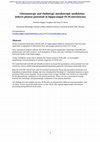
Oriens-lacunosum moleculare (OLM) cells are hippocampal inhibitory interneurons that have been im... more Oriens-lacunosum moleculare (OLM) cells are hippocampal inhibitory interneurons that have been implicated in regulation of information flow and synaptic plasticity in the CA1 circuit. Since anatomical evidence indicate that OLM cells express metabotopic cholinergic (mAChR) and glutamatergic (mGluR) receptors, such modulation of these cells may contribute to switching between functional modes of the hippocampus. Using a transgenic mouse line to identify the Chrna2-positive OLM cells, we investigated metabotropic neuromodulation of intrinsic properties of OLM cells. We found that both mAChR and mGluR activation increased the spontaneous action potential rate and caused the cells to exhibit long-lasting depolarizing plateau potentials following evoked spike trains. Both the mAChR-and mGluR-induced increased spontaneous firing rate and plateau potentials were dependent on intracellular calcium, and were eliminated by blocking Ca 2+-dependent transient receptor potential (TRP) cation channels. At the receptor level, Group I mGluRs were found to be responsible for the glutamatergic modulation of the plateau potentials. There was also a pronounced synergy between the cholinergic and glutamatergic modulation of the plateau potentials. Our findings provide insights into how OLM cells are modulated by different neurotransmitters, and are likely to have functional implications on how OLM cells regulate hippocampal information processing during different brain states. .

The Journal of Physiology, Nov 13, 2016
Kv2 channels underlie delayed-rectifier potassium currents in various neurons, although their phy... more Kv2 channels underlie delayed-rectifier potassium currents in various neurons, although their physiological roles often remain elusive. Almost nothing is known about Kv2 channel functions in medial entorhinal cortex (mEC) neurons, which are involved in representing space, memory formation, epilepsy and dementia. r Stellate cells in layer II of the mEC project to the hippocampus and are considered to be space-representing grid cells. We used the new Kv2 blocker Guangxitoxin-1E (GTx) to study Kv2 functions in these neurons. r Voltage clamp recordings from mEC stellate cells in rat brain slices showed that GTx inhibited delayed-rectifier K + current but not transient A-type current. r In current clamp, GTx had multiple effects: (i) increasing excitability and bursting at moderate spike rates but reducing firing at high rates; (ii) enhancing after-depolarizations; (iii) reducing the fast and medium after-hyperpolarizations; (iv) broadening action potentials; and (v) reducing spike clustering. r GTx is a useful tool for studying Kv2 channels and their functions in neurons.
Neuroscience Letters, Mar 1, 1987
Intracellular recordings were made from CAI pyramidal cells in rat hippocampal slices. Single act... more Intracellular recordings were made from CAI pyramidal cells in rat hippocampal slices. Single action potentials were elicited by injection of brief current pulses. Bath application of phorbol esters (4fl-phorbol-12,13-diacetate, 0.3 5 pM; or 4fi-phorbol-12,13-dibutyrate, 5 10/~M) broadened the action potential in each of the cells tested (n=9). The broadening reflected slowing of the repolarization, whereas the upstroke of the spike was unchanged. This effect may enhance transmitter release from synaptic terminals, and contribute to enhancement of synaptic transmission through activation of protein kinase C, a mechanism which has been associated with long term potentiation.

The Journal of Physiology, Feb 6, 2015
Kv7 (KCNQ/M) channels are known to control excitability and generate subthreshold M-resonance in ... more Kv7 (KCNQ/M) channels are known to control excitability and generate subthreshold M-resonance in CA1 hippocampal pyramidal cells, but their properties and functions have not previously been compared along the dorsoventral (septotemporal) axis r We used whole-cell recordings to compare electrophysiological properties of dorsal and ventral CA1 pyramidal cells in hippocampal slices from 3-to 4-week-old rats r Blockade of Kv7/M-channels with 10,10-bis(4-pyridinylmethyl)-9(10H)-anthracenone dihydrochloride (XE991) had a stronger impact on electrical properties in dorsal than ventral pyramidal cells, including input resistance, temporal summation, M-resonance, spike threshold, medium after-hyperpolarization, excitability, and spike frequency adaptation. r Voltage-clamp recordings revealed a larger amplitude and left-shifted voltage dependence of XE991-sensitive current (I M) in dorsal vs. ventral cells. r I M-dependent differences in excitability and resonance may be important for rate and phase coding of CA1 place cells along the dorsoventral axis and may enhance epileptiform activity in ventral pyramidal cells.

Nature Neuroscience, Dec 19, 2004
M channels are voltage-gated K + channels that underlie the non-inactivating M current (I M) in n... more M channels are voltage-gated K + channels that underlie the non-inactivating M current (I M) in neurons 1,2. These channels are of particular interest because they activate at membrane potentials that are more negative than the action-potential threshold and at which few other ion channels are active. Therefore, they may exert pivotal control over neuronal excitability and response patterns 1-4. This idea is supported by the finding that mutations in the human KCNQ2 and KCNQ3 genes, which are generally believed to encode the main M-channel subunits, cause a dominantly inherited form of human generalized epilepsy called benign familial neonatal convulsions (BFNC) 5. The BFNC phenotype is characterized by frequent seizures starting in the first week of life. In most cases, the seizures spontaneously disappear within weeks. Although BFNC is generally considered to be a benign epilepsy syndrome, the risk of recurring seizures in adolescence or adulthood is approximately 10-16%, and there are some reports of unfavorable outcomes, notably epileptic encephalopathy or mental retardation 6-8. In addition to the BFNC phenotype, a family carrying a KCNQ2 mutation also has myokymia, suggesting a potential role for KCNQ2 in motor-neuron or motor-axon function 9. The M current was first observed in frog sympathetic ganglia, in which it can be suppressed by muscarinic acetylcholine-receptor activation (hence its name I M) 1. Subsequently, M currents were recorded in mammalian brain neurons, including pyramidal cells of the hippocampus, where M channels are modulated by several neurotransmitters and neuromodulators 3,10. Several lines of evidence indicate that certain members of the KCNQ gene family encode M-channel subunits that may form homo-or heteromeric M channels 5. Heteromeric KCNQ2/KCNQ3 channels studied in in vitro expression systems have electrophysiological and pharmacological properties characteristic of M channels 11,12. In rat sympathetic ganglia, native M channels seem to be composed largely of KCNQ2/KCNQ3 subunits 13 , and these two subunits can be co-immunoprecipitated from human brain lysates 14. M channels in the mammalian brain may also contain KCNQ5 subunits 15. Immunohistochemical studies show overlapping KCNQ2 and KCNQ3 protein-expression patterns. This includes co-localization on both the somata and the dendrites of pyramidal and polymorphic neurons in the hippocampus and cerebral cortex, and co-localization on the somata of parvalbumin-positive hippocampal interneurons 16. KCNQ2/KCNQ3 subunits are also detected in axon initial segments and in central and peripheral axons 16,17. M-channel activity is thought to mediate neuronal excitability control and early spike-frequency adaptation 1,2,18,19 , and to generate afterhyperpolarizations of medium duration (mAHPs) during and after repetitive neuronal discharge 4,18. Thus, M-channel activity tends to stabilize the membrane potential, thereby preventing spiking. Accordingly, attenuation of M-channel activity through activation of G-protein-coupled receptors may enhance neuronal responses to excitatory input 2,10,18,20. However, the exact function of M channels in the brain-in particular their functions in animal behavior-as well as their roles in the development of epilepsy are not well understood. To examine the cellular, network and behavioral consequences of M-channel deficiency, we suppressed M currents in the mouse brain. Because genetic KCNQ2 gene deletions are lethal 21,22 , we generated mice expressing a KCNQ2 subunit with a dominant-negative pore mutation that can suppress M-channel activity by co-assembling with native KCNQ subunits. By using the Tet-Off system 23 and the prion-protein

The Journal of Physiology, Dec 1, 2002
Coherent network oscillations in the brain are correlated with different behavioural states. Intr... more Coherent network oscillations in the brain are correlated with different behavioural states. Intrinsic resonance properties of neurons provide a basis for such oscillations. In the hippocampus, CA1 pyramidal neurons show resonance at theta (θ) frequencies (2-7 Hz). To study the mechanisms underlying θ-resonance, we performed whole-cell recordings from CA1 pyramidal cells (n = 73) in rat hippocampal slices. Oscillating current injections at different frequencies (ZAP protocol), revealed clear resonance with peak impedance at 2-5 Hz at ≈33 °C (increasing to ≈7 Hz at ≈38 °C). The θ-resonance showed a U-shaped voltage dependence, being strong at subthreshold, depolarized (≈-60 mV) and hyperpolarized (≈-80 mV) potentials, but weaker near the resting potential (-72 mV). Voltage clamp experiments revealed three non-inactivating currents operating in the subthresold voltage range: (1) M-current (IM), which activated positive to -65 mV and was blocked by the M/KCNQ channel blocker XE991 (10 μm); (2) h-current (Ih), which activated negative to -65 mV and was blocked by the h/HCN channel blocker ZD7288 (10 μm); and (3) a persistent Na+ current (INaP), which activated positive to -65 mV and was blocked by tetrodotoxin (TTX, 1 μm). In current clamp, XE991 or TTX suppressed the resonance at depolarized, but not hyperpolarized membrane potentials, whereas ZD7288 abolished the resonance only at hyperpolarized potentials. We conclude that these cells show two forms of θ-resonance: ‘M-resonance’ generated by the M-current and persistent Na+ current in depolarized cells, and ‘H-resonance’ generated by the h-current in hyperpolarized cells. Computer simulations supported this interpretation. These results suggest a novel function for M/KCNQ channels in the brain: to facilitate neuronal resonance and network oscillations in cortical neurons, thus providing a basis for an oscillation-based neural code.
bioRxiv (Cold Spring Harbor Laboratory), Jul 16, 2022
Highlights Anesthesia caused a reduction in the perturbational complexity index (PCI ST) at bot... more Highlights Anesthesia caused a reduction in the perturbational complexity index (PCI ST) at both global (inter-areal) and local (intra-areal, across layers) cortical scales. Deep cortical layers (L6 and L5), exhibited strong connectivity with remote cortical areas during wakefulness but not during anesthesia. Layer 6 showed the strongest modulation of spike firing and spike field coherence compared to more superficial layers during wakefulness.

Nature, Dec 1, 1988
vation, others and especially antiprogestins forming abortive complexes with receptor and HREs. T... more vation, others and especially antiprogestins forming abortive complexes with receptor and HREs. The method described here may now be used to rapidly assign new compounds obtained by synthesis to each of these categories. The exact mechanisms of receptor interaction with HREs and susbequent changes in the rate of transcription are not understood. They may involve receptor dimerization or polymerization, interaction with transcription factors or RNA polymerase itself. Any one of these later events may be prevented from occuring when the receptor has bound an antagonist instead of an agonist. Analysis of transcription complexes formed in the presence of agonist and antagonist-receptor complexes should give insight into the mechanisms of hormone action following receptor binding to HREs. We thank D. Philibert (Roussel Uclaf) for the gift of RU486, C. Carreaud and S. Sar for technical assistance. MMTV-CAT construction was kindly provided by G. Schlitz (Heidelberg) and plasmid PHCwt by A.C.B. Cato (Karlsruhe). The manuscript was typed by V. Francois and N. Malpoint.

Objective methods for distinguishing conscious from unconscious states in humans are of key impor... more Objective methods for distinguishing conscious from unconscious states in humans are of key importance for clinical evaluation of general anesthesia and patients with disorders or consciousness, as well as for consciousness research. The Directed Transfer Function (DTF)-a measure of effective connectivity-was recently shown to accurately classify patients undergoing anesthesia as conscious or unconscious based on 1-second segments of raw EEG. Here, we test the generalizability of the DTF-based classification algorithm as an objective measure of conscious experience during anesthesia and correlate it with a well-tested index of consciousness: the Perturbational Complexity Index (PCI). We reanalyzed EEG data from an experimental study in which 18 healthy volunteers were randomly assigned to one of three types of general anesthesia: propofol, xenon, and ketamine. EEG was recorded before and during anesthesia, and DTF was calculated from every 1-second segment of the EEG data to quantify the effective connectivity between channel pairs. This was used to classify the state of each participant as either conscious or unconscious, and the classifications were compared with the participant's delayed report of experience. Finally, classifications were correlated with the PCI values obtained in the original study. The DTF analysis yielded two distinct patterns of DTF-based connectivity that corresponded well to the participants' reports about subjective experience, and was sufficient to accurately classify the state of the participants as conscious or unconscious. The algorithm was more likely to classify participants as conscious in the awake state than during propofol and xenon anesthesia (p<0.05), but not during ketamine anesthesia (p>0.05). Furthermore, the DTF-based confidence of being classified as conscious was highly correlated with PCI (r 2 =0.48, p<0.05). The DTF-based measure distinguished between conscious and unconscious states during different forms of general anesthesia and wakefulness, and it correlates strongly with PCI. These results provide further support for the notion that effective connectivity measured between EEG electrodes can be used to distinguish between conscious and unconscious states in humans.
BMC Pharmacology, Jun 1, 2005
Frontiers in Human Neuroscience, Jan 10, 2023

PLOS ONE, Dec 31, 2013
Extracellular (EC) recordings of action potentials from the intact brain are embedded in backgrou... more Extracellular (EC) recordings of action potentials from the intact brain are embedded in background voltage fluctuations known as the ''local field potential'' (LFP). In order to use EC spike recordings for studying biophysical properties of neurons, the spike waveforms must be separated from the LFP. Linear low-pass and high-pass filters are usually insufficient to separate spike waveforms from LFP, because they have overlapping frequency bands. Broad-band recordings of LFP and spikes were obtained with a 16-channel laminar electrode array (silicone probe). We developed an algorithm whereby local LFP signals from spike-containing channel were modeled using locally weighted polynomial regression analysis of adjoining channels without spikes. The modeled LFP signal was subtracted from the recording to estimate the embedded spike waveforms. We tested the method both on defined spike waveforms added to LFP recordings, and on in vivo-recorded extracellular spikes from hippocampal CA1 pyramidal cells in anaesthetized mice. We show that the algorithm can correctly extract the spike waveforms embedded in the LFP. In contrast, traditional high-pass filters failed to recover correct spike shapes, albeit produceing smaller standard errors. We found that high-pass RC or 2-pole Butterworth filters with cutoff frequencies below 12.5 Hz, are required to retrieve waveforms comparable to our method. The method was also compared to spike-triggered averages of the broad-band signal, and yielded waveforms with smaller standard errors and less distortion before and after the spike.

Frontiers in Human Neuroscience, Oct 5, 2022
In the field of consciousness science, there is a tradition to categorize certain states such as ... more In the field of consciousness science, there is a tradition to categorize certain states such as slow-wave non-REM sleep and deep general anesthesia as "unconscious". While this categorization seems reasonable at first glance, careful investigations have revealed that it is not so simple. Given that (1) behavioral signs of (un-)consciousness can be unreliable, (2) subjective reports of (un-)consciousness can be unreliable, and, (3) states presumed to be unconscious are not always devoid of reported experience, there are reasons to reexamine our traditional assumptions about "states of unconsciousness". While these issues are not novel, and may be partly semantic, they have implications both for scientific progress and clinical practice. We suggest that focusing on approaches that provide a more pragmatic and nuanced characterization of different experimental conditions may promote clarity in the field going forward, and help us build stronger foundations for future studies.

Uploads
Papers by Johan Frederik Storm